 As populations across the western world age, the ability to tackle conditions like arthritis is set to be increasingly important. I’ve written previously about a smart patch that can detect the condition in wearers, but this is not the only work being done to provide early detection.
A team from the University of Cambridge have developed an algorithm that can monitor the joints of patients to observe the change in their condition. The work, which was described in a recently published paper, could change how the severity of arthritis is assessed.
The system is able to detect minute changes in arthritic joints that could provide clinicians with a much greater understanding of just how osteoarthritis develops and allow the effectiveness of treatments to be assessed more accurately, all without tissue samples being required.
Spotting osteoarthritis
Osteoarthritis is the most common form of arthritis, but there isn’t currently any cure for the condition, with the only treatment surgery for artificial joint replacement. It’s traditionally identified via an x-ray that highlights a narrowing of the space between the bones of a joint, which is caused by a loss of cartilage. This is a robust method, but the very nature of x-rays makes it very hard to detect subtle changes over time.
“In addition to their lack of sensitivity, two-dimensional x-rays rely on humans to interpret them,” the team say. “Our ability to detect structural changes to identify disease early, monitor progression and predict treatment response is frustratingly limited by this.”
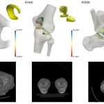
The new technique uses images collected from a standard CT scan. Such technology isn’t usually used to monitor joints, but does provide detailed images in 3D. The algorithm then analyzes these images using joint space mapping (JSM). It’s hunting for changes in the space between the bones of whatever joint is being analyzed.
Gold standard
The system was tested on a number of tests performed on human hip joints that had been donated for medical research. When the results were analyzed, it emerged that the system outperformed the current ‘gold standard’ procedure in terms of sensitivity.
“Using this technique, we’ll hopefully be able to identify osteoarthritis earlier, and look at potential treatments before it becomes debilitating,” the researchers say. “It could be used to screen at-risk populations, such as those with known arthritis, previous joint injury, or elite athletes who are at risk of developing arthritis due to the continued strain placed on their joints.”
The team believe that the technique could be a hugely valuable tool for the analysis of arthritis, whether in clinical or research settings. Not only does it provide accurate assessments and analysis, but by utilizing CT scans, it also exposes the patient to lower doses of radiation, meaning that it can be used more frequently for ongoing monitoring.