 A lot of the early forays of AI applications into healthcare have been around image processing. Companies such as IBM and DeepMind have both developed applications that process images in the hunt for illness and disease.
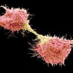
A recent paper from academics at Brown University have taken a similar approach to help support cancer research. The technique utilizes both microscopic imaging and machine learning to help scientists distinguish between two types of cancer cell that are strongly linked with tumor progression.
Spotting the transition
Central to the process is the epithelial-mesenchymal transition (EMT), which describes how docile epithelial cells turn into more aggressive, and therefore dangerous, mesenchymal cells. Tumors with more of this latter type of cell are significantly more malignant and harder to treat.
“We know that there are these different cell types interacting within tumors and that therapeutics can target these cells differently,” the authors say. “We’ve developed a model that can pick out these cell types automatically and in an unbiased way. We think this could help us better understand how these different cell types respond to drug treatment.”
The two cells can often be distinguished by their unique shapes, but these distinctions can be very subtle in nature and therefore hard for humans to detect. As such, being able to train a computer to do the job can be a life saver.
Learning what to look for
The algorithm was trained on an epithelial cell line that is used as a model for human breast cancer. The cells were manipulated to encourage the cells to rapidly undergo EMT. They were imaged throughout this process to provide a dataset by which the algorithm could be trained on.
When the resulting algorithm was put through its paces, it was able to categorize cells correctly with an accuracy of over 92%. It was then put to work on cells that undergo EMT via a less routine path than those used in the training set. Despite this more challenging environment, the algorithm still performed as well as the initial attempt.
The researchers hope that in time their algorithm will become an effective way of screening cancer drugs for effectiveness.
“When we do initial lab testing of drugs, we put cells on a plate, apply the drug, and see what lives and what dies,” they say. “This could provide us with a more nuanced picture of the drug’s effects, and help us to see whether sub-lethal doses may prime cells for resistance.”
The study also presented an interesting insight into possible new areas of study. For instance, they found that a small subset of cells were incredibly difficult to categorize, which may suggest a new type of cell type that is neither epithelial or mesenchymal.
As is so often the case with interesting research, it therefore poses fresh questions to consider, but it does nonetheless highlight the possibilities for image processing to make significant improvements in disease detection in the coming years.