Source: Advanced MR Analytics AB
Yesterday I wrote about an interesting project that was using machine learning to better understand and process the kind of medical images produced by CT scans. As with most machine learning projects, the process was much more effective when the algorithm was given a lot of data to train itself with.
So projects like the recently launched scanning study from the UK Biobank are so very important. The £43m study aims to create the largest collection of scans for everything from dementia to cancer, arthritis to heart attacks.
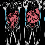
100,000 of those already signed up to the UK Biobank will have a series of scans of their heart, bones, brain, abdominal fat and carotid arteries to create an invaluable resource to improve health.
Rich data
These images will be analyzed alongside the huge database of medical information already collected from participants in the project. The data will available to all health scientists to access, and aims to give a fresh perspective on a huge range of conditions.
“This very large number of participants involved in the multimodal imaging study is impressive enough. But what makes it truly transformational is the opportunity to combine the rich imaging data with the wealth of other information already available or being collected from participants, particularly their health and diseases during follow-up for many years to come,” the team say.
A common problem with many medical studies is the relative shortage of data, and this huge repository promises to provide a valuable means of comparing and re-testing previous studies with this larger data source.
The new repository represents the largest brain imaging study ever conducted, providing valuable insight into a range of common conditions, whether dementia or motor neuron disease.
First steps
Some 8,000 people have already been scanned at the UK Biobank’s custom scanning facility in their Stockport headquarters, with this number set to be rapidly ramped up.
Participants will not receive any feedback on their health from the process unless something serious is uncovered. The images taken will include:
- MRI assessment of heart chamber diameter, the volume of blood flow, and how the heart changes as it pumps blood around the body, thickness of the heart wall and the size, shape and stiffness of the thoracic aorta, the vessel that delivers blood from the heart
- MRI measures of brain structure and function, volumes of grey matter and the mapping of major brain connections
- Dual-energy X-ray absorptiometry measures of bone density, osteoarthritic change at spine, hip and knee, fractures in the spine, and fat distribution throughout the body
- MRI measures of abdominal fat volume including in the liver and pancreas
- Ultrasound assessment of two major arteries, the carotid arteries, that run either side of the neck to the brain
It’s certainly a fascinating and worthwhile project, and with data such a precious commodity for the medical research industry, it’s one that will hopefully yield some very worthwhile results. Check out the video below for more information on the project.